Imaging
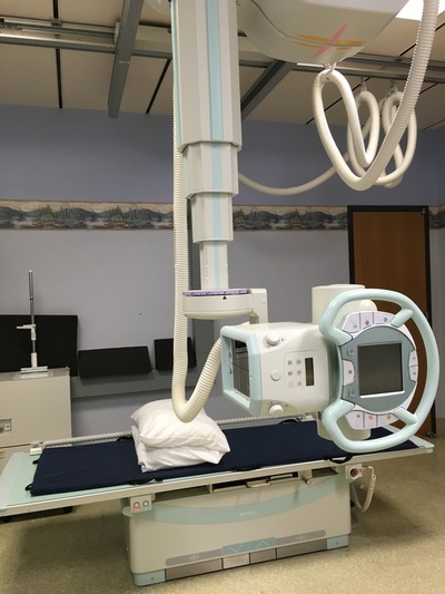
Our patients are given our time and attention and procedures are explained so you’ll know what to expect every step of the way. We also encourage our patients to ask questions if anything is unclear of if they want more information about a specific process or procedure. Petersburg Medical Center Imaging Department is capable of providing same or next day diagnostic testing in the following modalities:
INTERPRETATIONS:
All diagnostic imaging studies are interpreted by a team of radiologists from Radiology Associates PC (RAPC), in Eugene Oregon. RAPC allows PMC physicians access to the expertise of 20 sub-specialty-trained and supported radiologists via high-speed teleradiology. Images are stored on the RAPC digital Picture Archival & Communication system. PACS allows your digital images to be shared immediately and electronically with your physician. Interpretations are available within one hour for immediate requests, same or next day for the majority of our routine studies.
BILLING:
Patients will receive one bill from Petersburg Medical Center for the imaging study and another bill from RAPC for the Physician Interpretation.
STAFF:
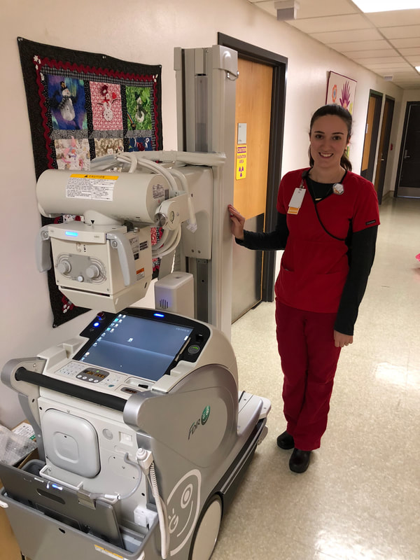
In addition, our Laboratory Staff are trained to provide diagnostic X-Ray.
HOURS OF OPERATION:
Monday – Friday, 8:00 – 5:00 p.m.
Closed observed holidays.
CT Coverage is 24/7/365 by On Call Technologist
IMAGING SERVICES:
Computed Tomography (CT)
Our Diagnostic Imaging department uses computed tomography, or CT to produce a series of detailed, three-dimensional images of parts of the body. The CT is a painless, fast scan that uses both special x-ray equipment and computers to produce images that can often provide more detailed information than regular x-rays.
Ultrasound
Ultrasound is a medical imaging technique that uses high-frequency sound waves similar to the sonar that dolphins and submarines use. Ultrasound imaging is used to study many of the body’s organs by using high frequency sound waves instead of radiation. When sound waves are recorded, they are immediately displayed on a monitor in real time. Although most individuals are familiar with an ultrasound as the device that allows them to see a picture of an unborn child, physicians also use ultrasound to see images and movement inside the patient’s body, including the body’s blood flow, and the size and function of many internal organs including the pancreas, liver, bladder and kidneys. Additionally, peripheral artery disease evaluations are available.
Mammography
Every woman has a better chance of fighting breast cancer if she understands the risk factors, how to detect the disease, and the options for treatment. The American Cancer Society recommends a screening mammography at age 40 and every other year thereafter until age 50. From ages 50 and older it is recommended to be screened yearly. For those with a strong family history of breast cancer: mother-sister diagnosed breast cancer, premenopausal, etc. please consult with your physician for guidelines about an earlier regimen.
PMC mammography is accredited by the American College of Radiology. PMC offers both Screening" annual mammograms and "Diagnostic" mammograms. If indicated, the diagnostic study may also include breast ultrasound. Specialized breast radiologists read and interpret the mammograms utilizing Computer-Assisted Detection (CAD) to aid in the earliest possible detection of breast cancer. If patients need a follow up study, this can often be performed the very next business day.
DEXA
Bone densitometry, also called dual-energy x-ray absorptiometry or DEXA, uses a very small dose of ionizing radiation to produce pictures of the inside of the body (usually the lower spine and hips) to measure bone loss. It is commonly used to diagnose osteoporosis and to assess an individual’s risk for developing fractures. DEXA is simple, quick and noninvasive. It’s also the most accurate method for diagnosing osteoporosis.
This exam requires little to no special preparation. Tell your doctor and the technologist if there is a possibility you are pregnant or if you recently had a barium exam or received an injection of contrast material for a CT or radioisotope scan. Leave jewelry at home and wear loose, comfortable clothing. You may be asked to wear a gown. You should not take calcium supplements for at least 24 hours before your exam.
Source: www.radiologyinfo.org
FREQUENTLY ASKED QUESTIONS:
How long does it take to get my results?
X-Rays performed at PMC are available to the physician immediately after the images are taken. The Radiologist report may take up to two days depending on the nature of the study.
Can my family come in for my OB Ultrasound?
We love to show you pictures of your baby! The decision to bring in family during or after the study is up to the discretion of the ultrasonographer, but usually after is best so the important diagnostic images are captured and this leaves plenty of time for your loved ones to see some great special views of your newest addition.
Can I get a copy of my report?
Yes, all you need to do is sign a Release of Information Form, and either you or someone you designate may pick up a copy of your report. Charges for CD images may apply.
Do I always need an appointment?
For X-Rays you do not need an appointment, but Ultrasound, Mammography and CT requires an appointment. We always do our best to get you in as soon as possible.
What do I need to do to prepare for my imaging study?
You may need a lab test to see if your kidneys are functioning properly for a CT contrast study. You may need to fast (nothing to eat or drink for 10 hours) or you may be asked to drink several glasses of water prior to the exam. When you schedule your appointment you will be given preparation instructions.
Why do I get two bills for my imaging studies?
One bill is for the study performed at Petersburg Medical Center (we take your image). The second bill is for the Radiologist Interpretation (they read your images) and this bill comes from RAPC. Please contact the Imaging Department for price quotes.
American College of Radiology www.acr.org
Radiology Associates PC www.rapc.com
- Diagnostic X-Ray
- General Ultrasound
- Obstetrical Ultrasound
- Vascular Ultrasound
- CT Scan
- Mammography
- DEXA Bone Density
INTERPRETATIONS:
All diagnostic imaging studies are interpreted by a team of radiologists from Radiology Associates PC (RAPC), in Eugene Oregon. RAPC allows PMC physicians access to the expertise of 20 sub-specialty-trained and supported radiologists via high-speed teleradiology. Images are stored on the RAPC digital Picture Archival & Communication system. PACS allows your digital images to be shared immediately and electronically with your physician. Interpretations are available within one hour for immediate requests, same or next day for the majority of our routine studies.
BILLING:
Patients will receive one bill from Petersburg Medical Center for the imaging study and another bill from RAPC for the Physician Interpretation.
STAFF:
- Sonja Ewing, RT (R), RDMS (Imaging Director)
- Tiffany Shelton, RDMS
- Elizabeth Thomas
In addition, our Laboratory Staff are trained to provide diagnostic X-Ray.
HOURS OF OPERATION:
Monday – Friday, 8:00 – 5:00 p.m.
Closed observed holidays.
CT Coverage is 24/7/365 by On Call Technologist
IMAGING SERVICES:
Computed Tomography (CT)
Our Diagnostic Imaging department uses computed tomography, or CT to produce a series of detailed, three-dimensional images of parts of the body. The CT is a painless, fast scan that uses both special x-ray equipment and computers to produce images that can often provide more detailed information than regular x-rays.
- What does a CT do? A CT scan can produce detailed images of organs, bones, soft tissues and blood vessels. CT scans are used to diagnose conditions such as cancer, musculoskeletal disease and trauma to certain areas of the body. The detailed results of a CT can eliminate or reduce the need for surgical biopsies and exploratory surgery.
- How to schedule a CT: Your physician will provide us with an order for CT examination and a member of our department will contact you to schedule an appointment. Patients under the care of a non – PMC physician must provide a written order to schedule an appointment. In some cases, pre-authorization is needed, depending upon insurance requirements. Sometimes you may be asked to prepare for the exam in advance by fasting or drinking a contrast agent.
Ultrasound
Ultrasound is a medical imaging technique that uses high-frequency sound waves similar to the sonar that dolphins and submarines use. Ultrasound imaging is used to study many of the body’s organs by using high frequency sound waves instead of radiation. When sound waves are recorded, they are immediately displayed on a monitor in real time. Although most individuals are familiar with an ultrasound as the device that allows them to see a picture of an unborn child, physicians also use ultrasound to see images and movement inside the patient’s body, including the body’s blood flow, and the size and function of many internal organs including the pancreas, liver, bladder and kidneys. Additionally, peripheral artery disease evaluations are available.
- How to Schedule an Ultrasound: Your physician will provide us with an order for an ultrasound examination and a member of our department will contact you to schedule an appointment. Patients under the care of a non – PMC physician must provide a written order to schedule an appointment. In some cases, pre-authorization is needed, depending upon insurance requirements. Sometimes you may be asked to prepare for the exam in advance by fasting or drinking a certain volume of water.
Mammography
Every woman has a better chance of fighting breast cancer if she understands the risk factors, how to detect the disease, and the options for treatment. The American Cancer Society recommends a screening mammography at age 40 and every other year thereafter until age 50. From ages 50 and older it is recommended to be screened yearly. For those with a strong family history of breast cancer: mother-sister diagnosed breast cancer, premenopausal, etc. please consult with your physician for guidelines about an earlier regimen.
PMC mammography is accredited by the American College of Radiology. PMC offers both Screening" annual mammograms and "Diagnostic" mammograms. If indicated, the diagnostic study may also include breast ultrasound. Specialized breast radiologists read and interpret the mammograms utilizing Computer-Assisted Detection (CAD) to aid in the earliest possible detection of breast cancer. If patients need a follow up study, this can often be performed the very next business day.
- How to Schedule a Mammogram: Women may self refer for an annual mammogram and PMC offers a "Birth Month" special. There is s special discount for screenings performed during your birth month. Diagnostic studies require physician order, either referred by the interpreting radiologist or the patient’s personal physician. Call 907-772-4291 ext 5755 to schedule a mammogram.
DEXA
Bone densitometry, also called dual-energy x-ray absorptiometry or DEXA, uses a very small dose of ionizing radiation to produce pictures of the inside of the body (usually the lower spine and hips) to measure bone loss. It is commonly used to diagnose osteoporosis and to assess an individual’s risk for developing fractures. DEXA is simple, quick and noninvasive. It’s also the most accurate method for diagnosing osteoporosis.
This exam requires little to no special preparation. Tell your doctor and the technologist if there is a possibility you are pregnant or if you recently had a barium exam or received an injection of contrast material for a CT or radioisotope scan. Leave jewelry at home and wear loose, comfortable clothing. You may be asked to wear a gown. You should not take calcium supplements for at least 24 hours before your exam.
Source: www.radiologyinfo.org
FREQUENTLY ASKED QUESTIONS:
How long does it take to get my results?
X-Rays performed at PMC are available to the physician immediately after the images are taken. The Radiologist report may take up to two days depending on the nature of the study.
Can my family come in for my OB Ultrasound?
We love to show you pictures of your baby! The decision to bring in family during or after the study is up to the discretion of the ultrasonographer, but usually after is best so the important diagnostic images are captured and this leaves plenty of time for your loved ones to see some great special views of your newest addition.
Can I get a copy of my report?
Yes, all you need to do is sign a Release of Information Form, and either you or someone you designate may pick up a copy of your report. Charges for CD images may apply.
Do I always need an appointment?
For X-Rays you do not need an appointment, but Ultrasound, Mammography and CT requires an appointment. We always do our best to get you in as soon as possible.
What do I need to do to prepare for my imaging study?
You may need a lab test to see if your kidneys are functioning properly for a CT contrast study. You may need to fast (nothing to eat or drink for 10 hours) or you may be asked to drink several glasses of water prior to the exam. When you schedule your appointment you will be given preparation instructions.
Why do I get two bills for my imaging studies?
One bill is for the study performed at Petersburg Medical Center (we take your image). The second bill is for the Radiologist Interpretation (they read your images) and this bill comes from RAPC. Please contact the Imaging Department for price quotes.
American College of Radiology www.acr.org
Radiology Associates PC www.rapc.com


